Thursday, 31 May 2012

Bat rabies virus evolves at rate X....or does it?

ResearchBlogging.org

One of the most interesting aspects of virology is the obligatory dependence of the virus upon its host. This dependence means that viruses have an intricate relationship with their host and have evolved to utilise the components of a specific host, although many viruses are happy to replicate in other organisms. For some it’s a necessity; arboviruses such as West Nile virus (WNV) must replicate in both vertebrate and invertebrate (in the case of WNV a mosquito) hosts to complete their lifecycles. Inevitably, this means that viruses can jump the species barrier leading to new (and potentially dangerous) scenarios where viruses behave differently in the new host. HIV for instance is thought to originally have crossed the species barrier into humans from chimpanzees.
Different hosts means different evolutionary pressures on a virus; factors such as bottle-necks and rates of transmission as well as immunological pressure will all impact upon the virus. A recent paper in PLoS Pathogens looks at how rabies virus evolves at different rates in different species of bat, and how the biology of bats impacts the subsequent evolution ofthe virus.


It seems that, once a virus is established in a particular species, the evolutionary rate remains fairly constant, although some virus lineages were able to evolve at a rate 5-22 times greater than that of others. Whilst the virus lineages themselves can show variation, they looked at whether there was variation in the rates of evolution in different species of bats and their ecology. Interestingly, the slower evolving lineages tended to be those evolving in bats of temperate zones, where the viruses were found to evolve at a rate nearly four-fold lower than that in tropical and sub-tropical areas. It seemed that the rate of evolution correlated not with a particular bat, but with where the bat was living. So do colder temperatures mean a lower metabolic rate and thus a reduced virus evolutionary rate? Apparently not. It appears it’s probably more to do with transmission rates and the impact that this has upon virus evolution.

Rabies virus evolves slower in species of bat dwelling in temperate areas

This all reminds me of a paper last year showing that different West Nile virus genomes predominated in different hosts (vertebrate vs. invertebrate) and that this affected the fitness of the virus in the alternative host (1). All in all, these kinds of study show just how complex viral evolution can be; can this all be explained by an evolutionary model based upon the concept of quasispecies?


(1) J. Virol. 2011. 85(23)12605-13

Streicker DG, Lemey P, Velasco-Villa A, & Rupprecht CE (2012). Rates of viral evolution are linked to host geography in bat rabies. PLoS pathogens, 8 (5) PMID: 22615575

Thursday, 24 May 2012

Learning from (virus induced) mistakes


One of my favourite popular science books is ‘Mutants: On Genetic Variety and the Human Body’. In the book Armand Leroi describes various humans which, although at the time may have ended up in one of those infamous ‘freak shows’, have a selection of different deformities of varying severity. The book is illuminating in that it follows the idea that if we can see what made something go wrong during development, then it tells us something about what happens when things go to plan. Knowing what we do now about development, it is possible to look back at these historic cases and explain them biologically. For instance, if someone is born with a deformed limb, and we find that they have a mutation in a specific gene, we can say that the gene is somehow involved in limb development.

Mutations are natural. As Leroi sates, “we are all mutants, but some of us are more mutant than others”. However, they can also be induced. Studies of embryogenesis and development are inextricably linked with Drosophila fruit flies. Many facets of development were figured out by inducing mutations in Drosophila with X-rays, resulting in a bewildering number of mutants being described with diverse and imaginative names. Individual pathways and genes have since been correlated with particular phenotypes.

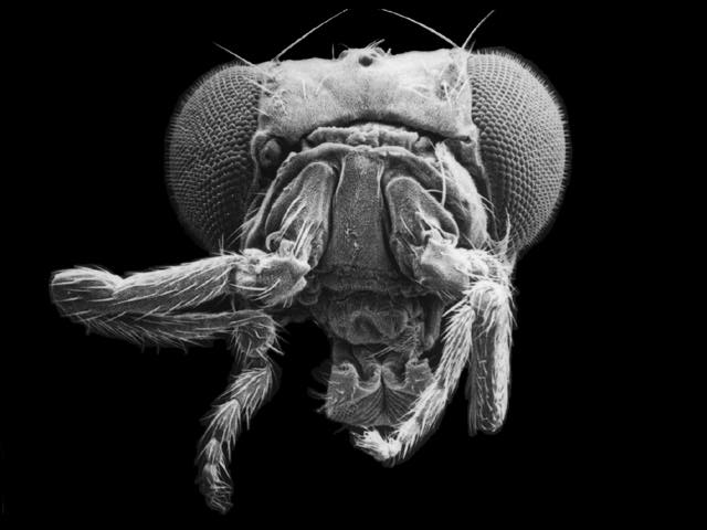
Antennapedia mutant of Drosophila: flies possess legs in place of antennae

Not all errors in development are directly linked to mutations. 1957-1961 saw the tragic incident of thalidomide, a drug taken by pregnant women to ease the discomfort of morning sickness. It soon became apparent that thalidomide was teratogenic with the resulting offspring sometimes possessing, among other deformities, severely stunted limbs. Clearly the presence of thalidomide interferes with foetal development. Although it is still not fully understood, various ideas exist about the way in which it causes the effects, including angiogenesis in the developing limb, thus stunting outgrowth (1).

A mother sheep sniffs her Schmallenberg virus-infected offspring (Kreisverwaltung)
A ewe investigates her Schmallenberg-affected lamb
When all of the images of lambs with arthrogryposis started to emerge a few months ago as a result of Schmallenberg virus infection during gestation, it reminded me of the approach ‘Mutants’ was following. As a Bunyavirus, Schmallenberg virus only has 6 proteins. By looking into the pathways which these proteins disrupt it could be possible to learn something about proper limb development. Whilst looking into this a bit more, a paper has been published detailing the infection of foetuses with Cache Valley virus (2), another Bunyavirus which also causes congenital abnormalities in sheep.

The paper looks at the localisation of virus infection and the time-frame of infection and subsequent development of the malformations. The data support previous work revealing that infection of the central and peripheral nervous systems is responsible for reduced foetal movement and the eventual development of the arthrogryposis. This confirms the idea of using viruses to investigate development: observation of the tropism of the virus and correlating this to the deformities of the resulting offspring allows, at a somewhat macroscopic level, identification of the role of particular tissues in development. As more and more methods are developed it may be possible to elucidate the molecular pathways which are disrupted by infection with these viruses, thus linking development to a specific pathway. Clearly the results are likely to be more ‘pointers’ of what’s happening as opposed to precisely defined interactions, but learning from mistakes is always a good thing!

(1) Proc Natl Acad Sci USA. 2009. 106(21):8573-8.
(2) J. Virol. 2012. 86(9):4793-800

Thursday, 17 May 2012

FMD 2001....rise of the “self-styled experts"?


Whilst searching through a pile of papers recently I came across one I recalled as being refreshingly open about its message. The authors, advocates of traditional approaches for the control of Foot and Mouth Disease (FMD), were suggesting that, as useful as models may be, they were not necessary in the control of the FMD outbreak in the UK in 2001. Indeed, it was speculated that the implementation of models may have exacerbated the problems.


Traditional FMD control has and continues to revolve around a stamping-out approach, supplemented by increased biosecurity, surveillance and the implementation of control zones. If an animal on a farm is found to have FMD, the farm becomes an ‘infected premises’ (IP) and all FMD-susceptible species on the farm are slaughtered. Other premises with epidemiological links to the IP are then classed as ‘dangerous contacts’ (DC) and also have susceptible stock removed.

The controversial aspect - the contiguous cull - in the 2001 outbreak was that, rather than having identified epidemiological links, a farm was classed as a DC simply by having land contiguous to an IP. The contiguous cull was deeply unpopular and was largely supported by modelling. It appears to be this point which irked the authors most; their view was that FMD policy during the epidemic was now being influenced by “self-styled ‘experts’” sat behind computers producing models generated using data from previous outbreaks with different strains of virus, inaccurate livestock census data, and ‘inaccurate biological assumptions’.

Location of the FMD IPs in the UK in 2001.
From Mansley et al 2011


Is this fair enough? Hindsight is a wonderful thing and models can easily be criticised for being unable to capture the complexity of an outbreak situation. Historically models were reductionist by their nature, but science is not static. The emergence of novel modelling approaches, along with the technological advances allowing the collation of more detailed quantitative and spatial datasets, as well as increasingly powerful computers, results in models of ever more sophistication and precision. The power of models to unveil patterns of spread and illuminate the most effective ways in which to employ control measures cannot, and will not, be ignored. Is this not the best way forward?

This is not a new debate; for every paper extolling the virtues of traditional approaches (if it ain’t broken....) there will be another detailing the benefits which can be reaped from models. An amalgamation of the two would appear to be the way forward. It would appear that models are here to stay, with the H1N1 pandemic influenza outbreak a classic example. So now there are new questions to ponder; ultimately, who should have the final say?

So if an outbreak happens tomorrow what should happen? I leave that for you to decide.

Wednesday, 9 May 2012

"What are you looking at?"


Not just a phrase favoured by self conscious teenagers, “what are you looking at?” is a question that anyone should be asking when trying to detect something. Diagnostics is a classic case.

With the rise of modern molecular approaches, traditional virological assays would appear to now languish in the dark shadows cast by the likes of real-time RT-PCR and next generation sequencing. This is, of course, unsurprising; molecular assays have many advantages such as speed and sensitivity relative to traditional virological methods. Molecular assays have, and continue to, revolutionise diagnostic capabilities in a multitude of ways. One of the biggest impacts is when unknown viruses arrive. The outbreak of SARS was first reported in March 2003; by May that year a virus had been isolated and, via sequencing, had been identified as a coronavirus. Most recently Schmallenberg virus was identified using metagenomic sequencing of a blood sample from a clinically affected cow. In terms of disease though, Koch’s postulates cannot be fulfilled without first isolating the suspected agent...

Pathogen detection in animals can be divided into either direct detection of the pathogen, or detection of evidence that the pathogen has been there. The latter generally refers to the detection of an immune response, most commonly antibodies. Detection of the pathogen can be further divided into detection of genome, protein or, in the case of viruses, by isolation of the virus. They may sound similar, but in reality it’s not. When it’s stated that a virus was detected by PCR, it can be argued that live virus was not being detected, but actually only a fragment. This may sound trivial, and in many cases PCR positive and virus positive equate, but it is of significance if say an unknown species of blood-sucking insect is responsible for spreading a virus from animal to animal. Any insect that has fed on a viraemic host can potentially be positive for the virus according to PCR, regardless of whether it is capable of supporting virus replication, or even if the virus is still ‘live’. We found a similar scenario with Foot and Mouth Disease Virus - some samples which were negative for live virus were positive by ELISA and/or real-time RT-PCR (detecting the proteins or genome respectively of viruses that had fallen apart in acidic conditions and were no longer infectious). What does this mean regarding whether an animal is infectious? Assuming the absence of contamination, and in the case of FMDV, genome in a clinical sample probably means the animal was infectious. No virus, no genome. On the other hand, it is well established for Bluetongue virus that animals can frequently be positive for RNA by RT-PCR, but non-infectious for biting midges.  
  
And characterisation? Does a PCR assay suggesting a virus is of serotype X categorically mean it is a virus with serotype X? Well, probably. But whilst a PCR assay detects the genomic sequence, a serotype reflects the expressed form of the genome, i.e. the phenotype. Perhaps this is pedantic, but chimpanzees are 95-99% identical to humans – a PCR assay might easily suggest they are the same, but ultimately they are different. Serotype classically refers to protection against an identical serotype, which by definition refers to serum, i.e. antibodies, and antibodies generally recognise protein.  The advent of sequencing has increased the precision of this and now the aim is to combine the two using mathematical modelling such that serotype is accurately predicted by the genomic sequence.

In the future, virus diagnostics may well involve simply sequencing everything in a sample, resulting in the full spectrum of pathogens present – the equivalent of a deep-sea trawler compared to fishing with a rod and some bait. As such metagenomics approaches expand an enticing prospect is the derivation of a prognosis based upon the molecular signatures present.

Ultimately though, if you’re interpreting diagnostics results, you need to know what you’re looking at!